02Measure the response: select the relevant omics layers
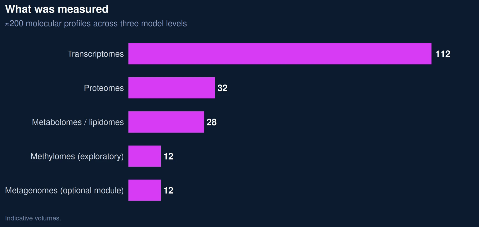
Skin Biology Xplorer draws on five omics layers.
CASE STUDY · SKIN BIOLOGY · HEALTHY AGING
How Skin Biology Xplorer combines 2D cells, reconstructed 3D skin and human skin explants with genomic, transcriptomic, proteomic, metabolomic and metagenomic readouts to characterize what an anti-aging ingredient does, through which biological pathways, and which claims the evidence can support, an illustrative case study.
Illustrative case study. This scenario shows how Skin Biology Xplorer can be applied to an anti-aging question. It does not describe a real client project or an existing ingredient: models, results and timelines are illustrative and depend on the active, the target claim and tissue availability. Data from cellular, 3D and explant models can support mechanistic claims; consumer-perceivable efficacy claims require studies in volunteers.
Download the full case study (11 pages, PDF)
In this case study, “skin longevity” refers to the long-term maintenance of biological functions associated with skin resilience, including redox homeostasis, extracellular-matrix integrity, cellular senescence control, epidermal homeostasis and energy metabolism.
2D cells, 3D skin or explants selected to match the question
Genomic, transcriptomic, proteomic, metabolomic and metagenomic readouts
A pathway is strengthened when independent layers converge
Mechanism of action and evidence for claims
A cosmetic ingredient supplier had developed a fermented botanical extract positioned around “skin longevity.” Early data, a procollagen ELISA and a chemical antioxidant assay, were encouraging, but they did not answer the questions raised by its customers’ regulatory and R&D teams.
The request: characterize what the extract does in skin, understand how it does it, and determine which claims the evidence can support, and which it cannot.
No single model answers every question. We built a model ladder in which each level addresses a different question, using the same comparators: vehicle, extract and retinol3, a well-documented anti-aging comparator. Aging biology was modeled in two ways: cellular senescence, one of the hallmarks of aging4, and UV-induced photoaging, a major contributor to visible skin aging5.
12D cells
Is the extract safe and active at cellular level? At which concentrations? Which pathways respond first?
5 non-cytotoxic concentrations + vehicle, n = 4; 4-week extended culture for epigenetic readouts
2Reconstructed 3D skin
Does the formulated active work after topical application across a living epidermis?
Non-irradiated control, UV + vehicle, UV + 3% extract, UV + 0.1% retinol; n = 4 tissues per arm; optional colonized version for microbiome studies
3Human skin explants
Does the effect persist in real, aged human skin despite donor-to-donor variability?
7-day culture, daily topical application; same four arms; epidermis and dermis analyzed separately
Skin Biology Xplorer draws on five omics layers.
Each data layer was first analyzed independently and then integrated. A biological pathway was considered strengthened when independent layers or models converged on the same direction of effect.
| Epigenomics | Transcriptomics | Proteomics | Metabolomics | Metagenomics | Phenotypic test | Conclusion | |
|---|---|---|---|---|---|---|---|
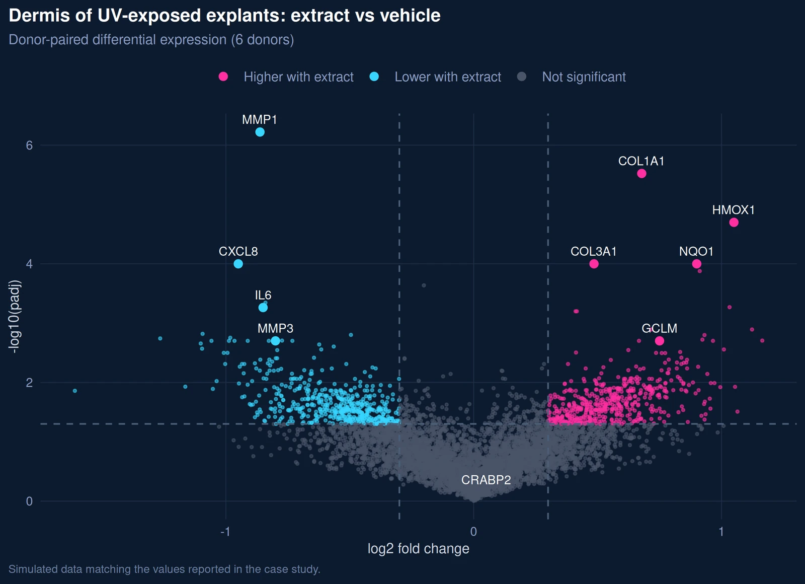
| Dermal matrix synthesis | – | ↑ | ↑ | – | – | ↑ | Supported |
| Matrix degradation (MMP-1) | – | ↓ | ↓ | – | – | – | Supported |
| Oxidative-stress response (NRF2) | – | ↑ | – | ↑ | – | ↓ | Supported |
| Senescence and SASP | – | ↓ | ↓ | – | – | ↓ | Supported |
| Energy metabolism (NAD⁺) | – | = | – | ↑ | – | – | In vitro only |
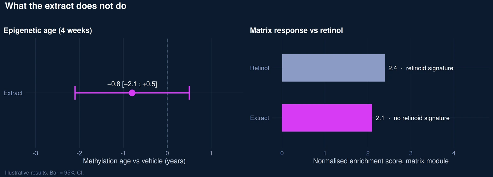
| Epigenetic age | = | – | – | – | – | – | Not supported |
| Microbial-community balance | – | – | – | – | = | – | Preserved (model) |
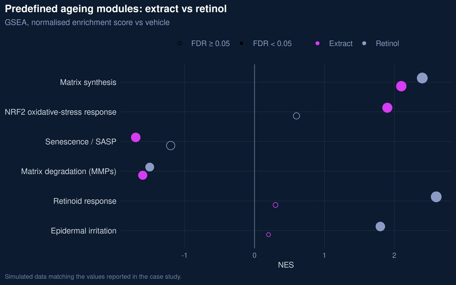
| Retinoid signaling | – | = | – | – | – | – | Not activated |
| Candidate claim (model wording) | Evidence | Conclusion |
|---|---|---|
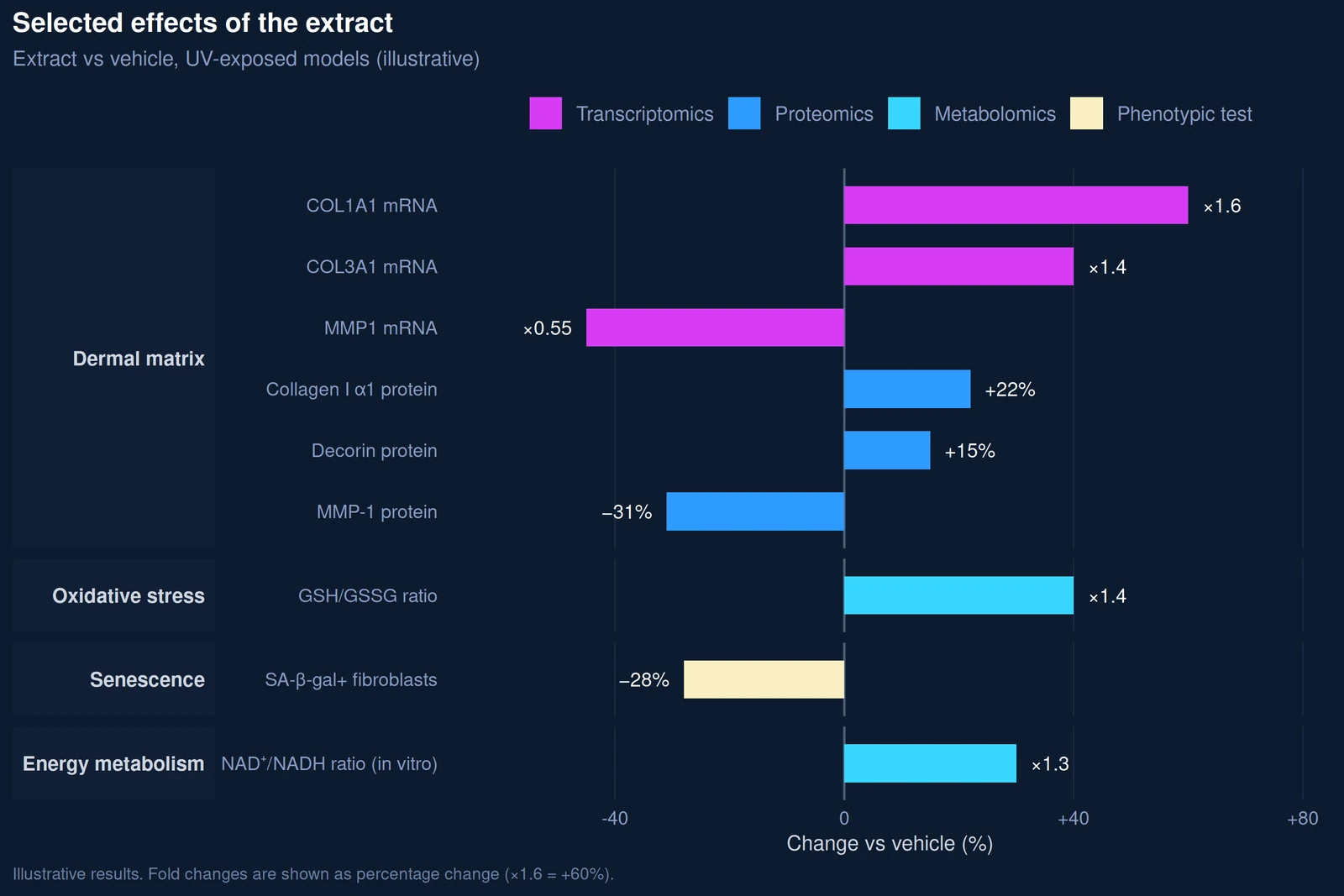
| “Helps preserve the dermal matrix of UV-exposed human skin (ex vivo)” | Collagen I and decorin ↑ at gene and protein levels; MMP-1 ↓; collagen staining ↑ | Supported: 3 layers, 2 models |
| “Helps protect skin cells against oxidative stress (in vitro, ex vivo)” | NRF2 targets ↑; GSH/GSSG ↑; oxidized lipids and ROS ↓ | Supported: 2 layers + phenotype |
| “Reduces markers of cellular senescence (in vitro)” | SA-β-gal ↓; SASP transcripts and secreted proteins ↓ | Supported |
| “Acts through a non-retinoid molecular profile” | No retinoid-response signature detected, unlike the retinol comparator | Supported: mechanistic positioning |
| “Supports cellular energy metabolism (in vitro)” | NAD⁺/NADH ↑ in fibroblasts only | Restricted: one layer, one model; wording strictly limited to in vitro |
| “Does not significantly alter the composition or functional profile of a skin microbial community in this model” | No statistically significant change in composition or function in colonized 3D skin | Restricted: model-specific; volunteer study required for an in vivo claim |
| “Reverses epigenetic / biological age” | −0.8 years; 95% CI −2.1 to +0.5 | Not supported: not claimed |
Under Commission Regulation (EU) No 655/2013, cosmetic claims must meet six common criteria, including evidential support and honesty. Laboratory models can substantiate mechanistic claims framed as in vitro or ex vivo findings; consumer-perceivable efficacy claims, such as visible wrinkle reduction, require studies in volunteers. Final claim wording remains the responsibility of the client’s regulatory team.
Have a similar question?
Tell us about your active ingredient, target claim and available models. We will propose an evidence plan, model ladder, omics layers and comparators, before processing a single sample.
Discuss your study